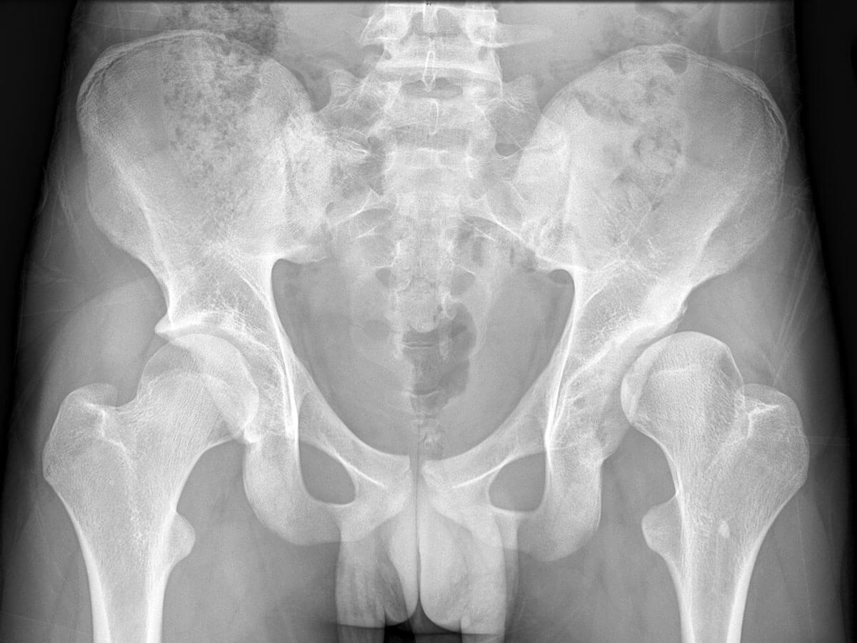
Left Acetabular Roof

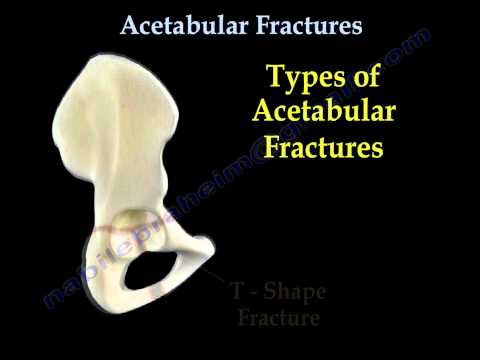
Acetabular Fracture Wikipedia
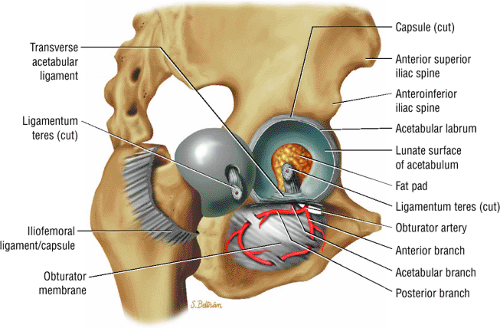
The Hip Musculoskeletal Key

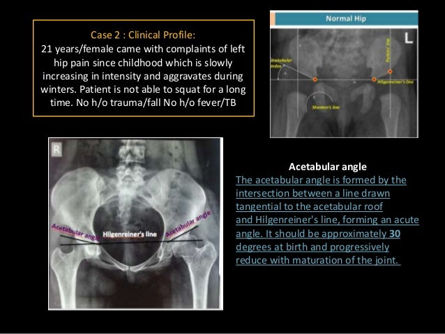
Acetabular Angle Radiology Reference Article Radiopaedia Org

Roof Arc Angle Of Matta The Roof Arc Orthopaedic Cases Discussion Facebook
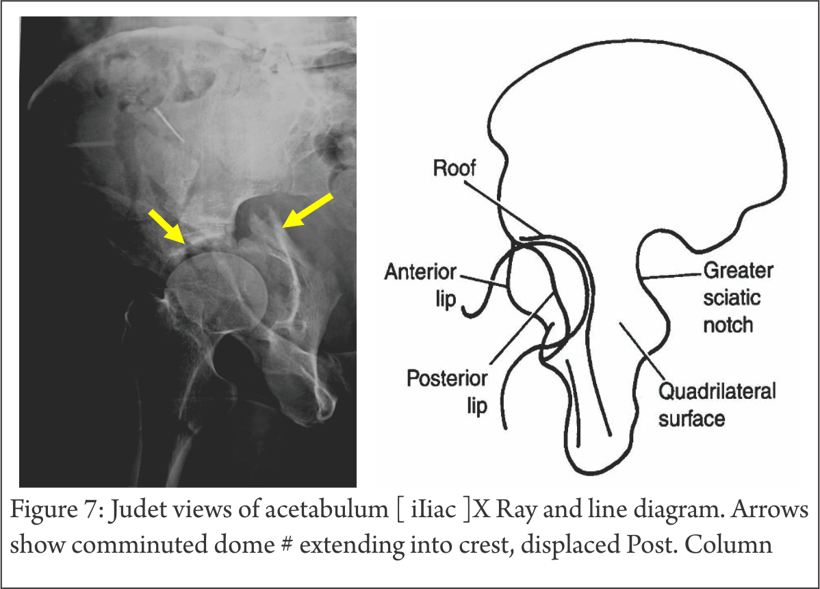
Understanding Clinical Radiology Of Fracture Acetabulum Trauma International
Acetabular Dysplasiar Orthopaedic Surgeon In Wantrina Melbourne Knox Orthopaedic Group

Fractures occur in a bimodal distribution.
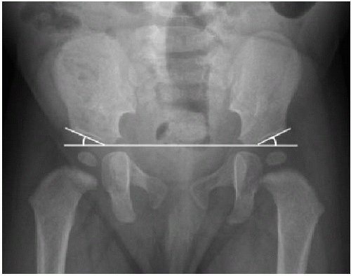
Left acetabular roof.

A D A Acetabular Dysplasia With A Short And Steep Acetabular Roof And Download Scientific Diagram
Http Links Lww Com Jbjs E539
Role Of Medical Imaging In Developemental Dysplasia Of Hip Dr Muhamma

Sketch Of The Three Main Types Of Acetabular Roof Dysmorphia A Download Scientific Diagram
Ddh Final Dt2

A Plain Radiograph Showing A Left Displaced Acetabular Anterior Wall Download Scientific Diagram
Developmental Dysplasia Of The Hip Radiology Reference Article Radiopaedia Org

Conventional Ap Radiograph Of Pelvis Shows Lytic Expansile Lesions Of Download Scientific Diagram

Figure 1 From Isolated Supra Acetabular Insufficiency Fracture A Case Report Semantic Scholar

Acetabular Fractures Radiology Key

Stress Fracture In Acetabular Roof Due To Motocross Case Report
Acetabulum Fracture Physiopedia

Figure 3 From Imaging Of Chondral Lesions Including Femoroacetabular Impingement Semantic Scholar

Measurement Of The Acetabular Index Ai From The Lateral End Of The Download Scientific Diagram

Acetabular Fracture Radiology Reference Article Radiopaedia Org

A D A This Ap Pelvic Radiograph Shows Acetabular Dysplasia Of The Download Scientific Diagram

Girl With Left Hip Pain Mechanical Symptoms
This Radiograph Documents The Measurement Of The Acetabular Roof Download Scientific Diagram
Https Encrypted Tbn0 Gstatic Com Images Q Tbn 3aand9gctohutsw4vrurdeistfd5zh9qok7nxgciawomkl0azqzgmadnzj Usqp Cau
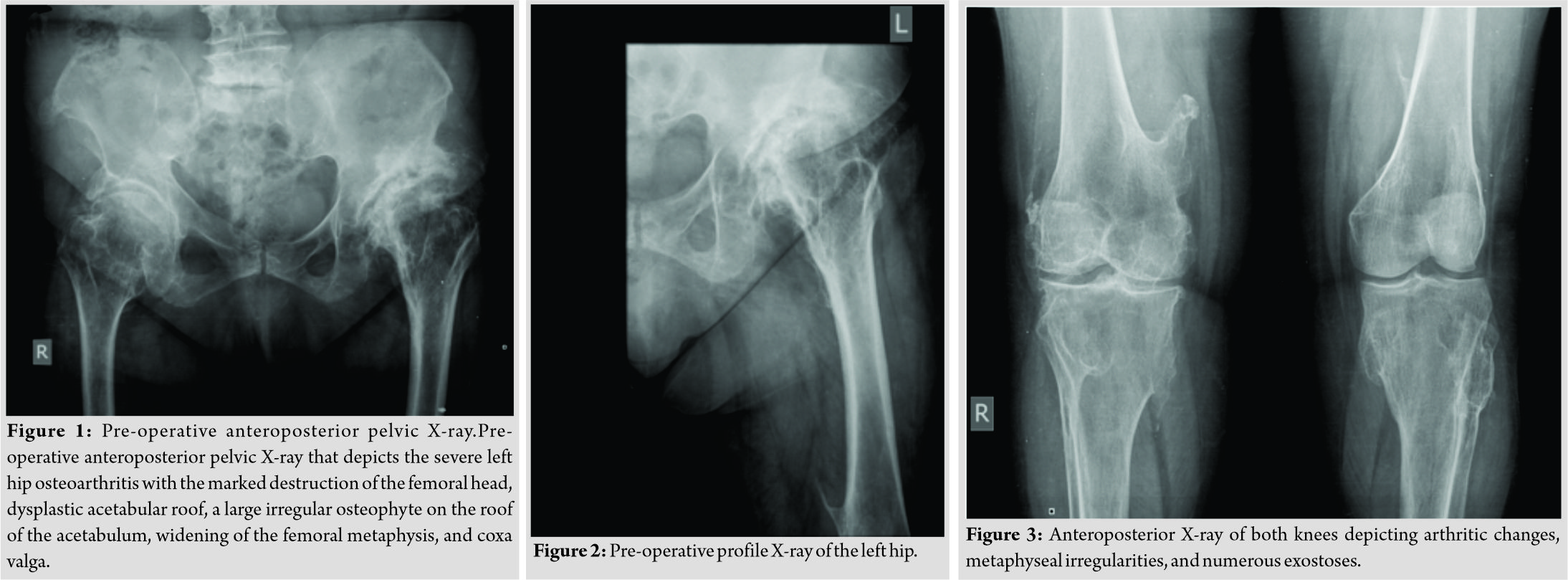
Technical Considerations Of Complex Primary Total Hip Arthroplasty In A Rare Case Of Combined Achondroplasia And Hereditary Multiple Exostosis Syndromes Journal Of Orthopaedic Case Reports

Human Anatomy Lab 2 Bones Of The Lower Body Flashcards Quizlet
Hip Disorders Radiology Key

A B A A 22 Year Old Female Patient With Bilateral Borderline Download Scientific Diagram

Primary Total Hip Arthroplasty After Acetabular Fracture By Dana C Mears And John H Velyvis J Bone Joint Surg Am Volume 82 9 September 1 Ppt Download
Source : pinterest.com
